By Mohamed Eldernawi, BDS, ALD member, ALD Fellowship
 One of the most difficult cases in restorative dentistry is the deep subgingival caries in Class II and Class V cavity preparations. The subgingival location of the caries makes it difficult to clean without trauma to soft tissue that will lead to bleeding which is difficult to control and sometimes causes a delay in treatment by postponing the restoration to another visit so the dentist can work in a dry field.
There are several clinical approaches to manage the soft tissue and expose the caries such as surgical gingivectomy by blade or by thermacut bur, however, this may lead to bleeding which is difficult to control at the same visit especially if the tissue was inflamed. This article proposes the use of a diode laser to manage the soft tissue without bleeding and expose the hidden caries under the gum so the dentist can finish the restoration with a clean dry field in the same visit. The laser technology is a useful noninvasive method alternative to the surgical technique by blade or thermacut bur. The laser technology also facilitates healing without side effects due to its bactericidal and biostimulation effects. The disadvantages of laser technology are the cost of the device, especially erbium lasers, and the need for the sufficient knowledge to use it safely and effectively.
This is a clinical case of laser-assisted deep subgingival Class V caries of an upper right canine with 2 weeks follow-up. Images follow.
 |
|
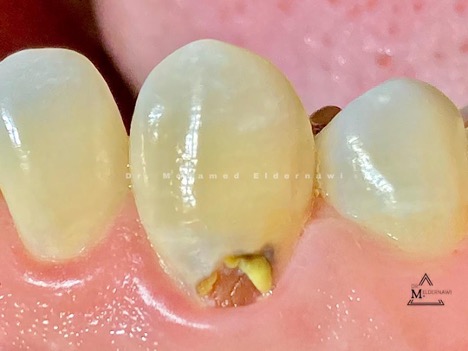
Fig. 1: Initial presentation of deep subgingival caries This is a short 63-second video showing laser-assisted gingivoplasty of the same case. |
 |
|
Fig 2: Immediate postoperative view after laser gingivoplasty |
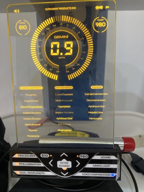 |
| Fig. 3: Gemini diode laser from Ultradent |
 |
|
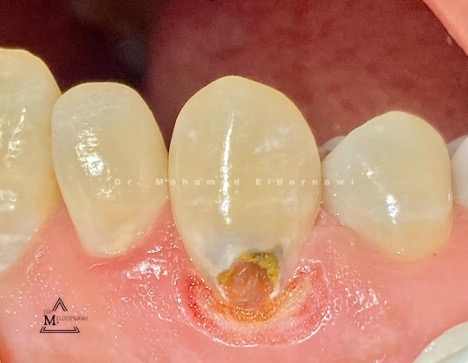
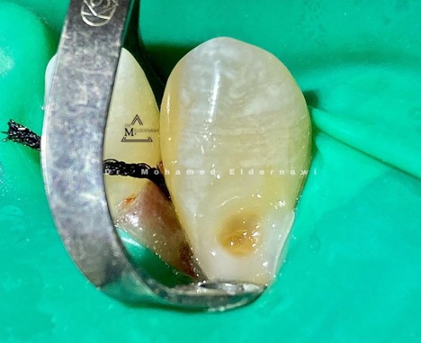
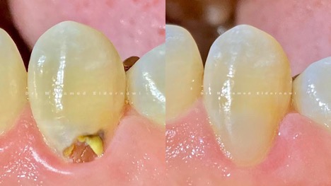
Fig. 4: Caries exposed with hemostatic effect on soft tissue (right image) |
 |
|
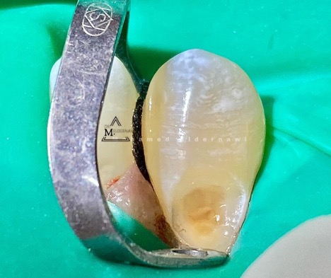
Fig. 5: Rubber dam isolation with #44 clamp from KSK and #000 retraction cord is placed for extra retraction. |
 |
|
Fig. 6: After selective etching and bonding, |
 |
|
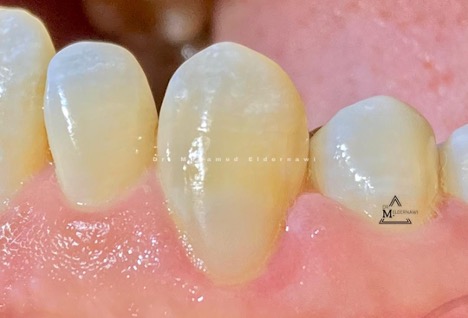
Fig. 7: Immediate postoperative view after finishing and polishing |
 |
|
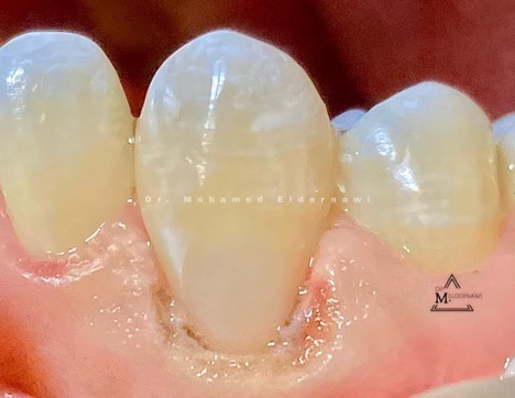
Fig. 8: 2-week follow-up showing healing of tissue around the restoration |
 |
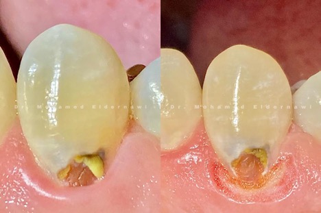
| Fig. 9: Before-and-after clinical pictures at 2 weeks |
Conclusion
Laser technology represents a useful method for soft tissue management in deep subgingival caries in Class V and Class II cases. Laser technology has a major impact in restorative dentistry due to the hemostatic effect that gives the clinician a chance of exposing the subgingival caries and finishing the restoration in the same visit. Also it provides greater patient satisfaction and motivation in the absence of bleeding during the procedure.